New microscopes to unravel the mysteries of brain organization
Collaboration
Researchers around the world share their stunning images and insights: The open source mesoSPIM Initiative
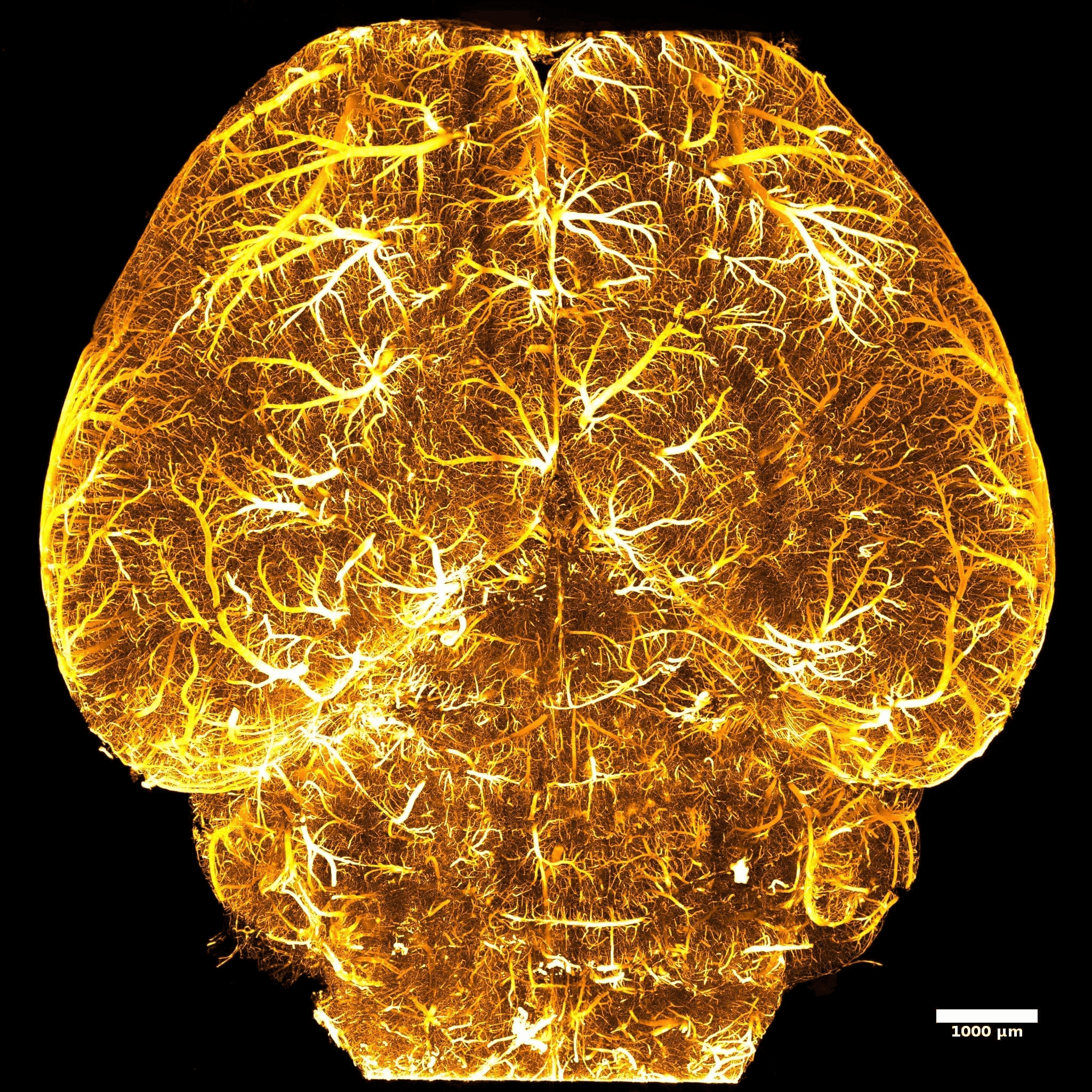
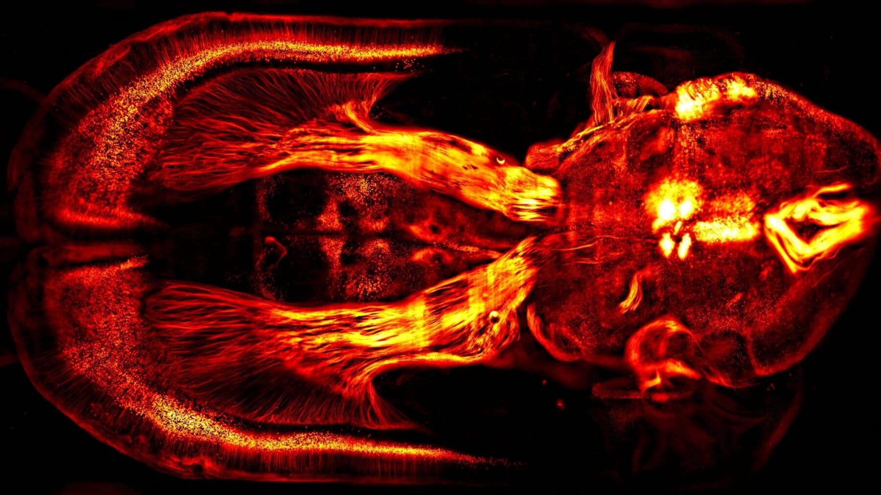
The secret of capturing exquisite brain images with a new generation of custom-built microscopes is revealed today in Nature Methods. The new microscopes, known as mesoSPIMs, can image the minute detail of brain tissue down to individual neurons that are five times thinner than a human hair, and can uncover the 3D anatomy of entire small organs, faster than ever before.
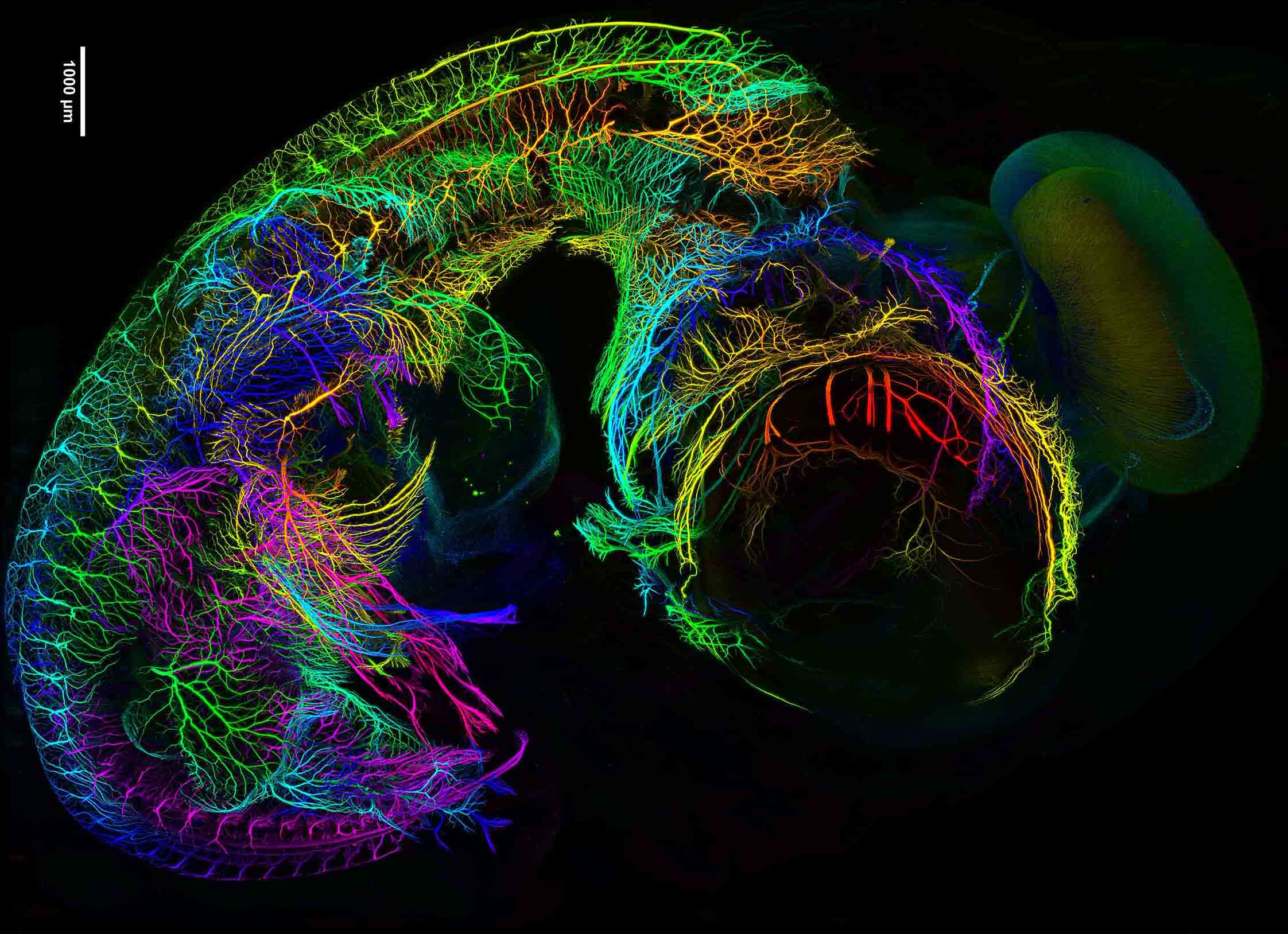
This large-scale dataset reveals the developing nervous system of a seven-day old chicken embryo captured with a mesoSPIM microscope. Credit: mesoSPIM.org
MesoSPIMs provide new insights into brain and spinal cord organization for researchers working to restore movement after paralysis or to investigate neuronal networks involved in cognition, pleasure, or drug addiction.
MesoSPIMs create high-resolution images of large samples faster than existing microscopes so are beneficial for rapidly screening many samples. A new open-source initiative, comprising top European researchers in neuroscience, is driving dissemination of mesoSPIMs globally by sharing their expertise and excitement as well as stunning images and videos.
MesoSPIMs, short for ‘mesoscale selective plane-illumination microscopes’, are light-sheet microscopes. Unlike traditional microscopy in which specimens are cut in slices with a blade before being viewed on a slide under a microscope, light-sheet microscopes optically slice samples with a sheet of light. This optical sectioning captures slivers of image without damaging the sample. The imaged slices are then combined to reconstruct a detailed three-dimensional image of a whole organ or specimen. The data sets produced by standard light-sheet microscopes are very large and analysing them is time consuming. MesoSPIMs get around this problem with innovative optical technologies that allow fast scanning as well as direct visualization and quantification of the captured data.
The mesoSPIM Initiative, started by Dr. Fabian Voigt in the group of Prof. Fritjof Helmchen at the Brain Research Institute, University of Zurich, enables the integration of cutting-edge technologies into research labs worldwide. “We created the open-source mesoSPIM Initiative to share the latest developments in microscope instrumentation and software with the imaging community. Anyone seeking high-quality anatomical data from large samples now has the information they need to build and operate their own mesoSPIM.” said Voigt.
The power of the initiative lies in the insights brought from differing disciplines, such as physics, developmental biology and neuroscience which allows microscope development, and brain research, to flourish.
There are currently seven mesoSPIMs in operation across Europe and several more instruments under construction. One of the new mesoSPIMs is hosted by the Advanced Lightsheet Imaging Center (ALICe) at the Wyss Center. Open to external users, ALICe offers a complete pipeline from sample preparation to image analysis, under the scientific guidance of experts from the University of Geneva and the École Polytechnique Fédérale de Lausanne (EPFL).
Dr Stéphane Pagès, ALICe Scientific Coordinator said: “We are very proud to have one of only seven mesoSPIM microscopes in the world at the Wyss Center. MesoSPIM microscopes solve the longstanding problem of how to achieve exceptional image quality in large samples over very short time-scales. We are delighted to be part of the initiative that brings this technology to the world.”
The mesoSPIM Initiative is aimed at research groups and imaging facilities with experience in building and supporting custom microscopes. A mesoSPIM can be installed in few days and typically requires a budget of around $200K.
‘The mesoSPIM initiative – open-source light-sheet microscopes for imaging cleared tissue’ is published in Nature Methods.
Play Video
This large-scale dataset reveals the developing nervous system of a seven-day old chicken embryo captured with a mesoSPIM microscope. Credit: Mesospim.org
A new model of cooperation and collaboration to accelerate translational research and development in the field of neurotechnology and artificial intelligence.