Take a fantastic 3D voyage through the brain with new immersive virtual reality system
Collaboration
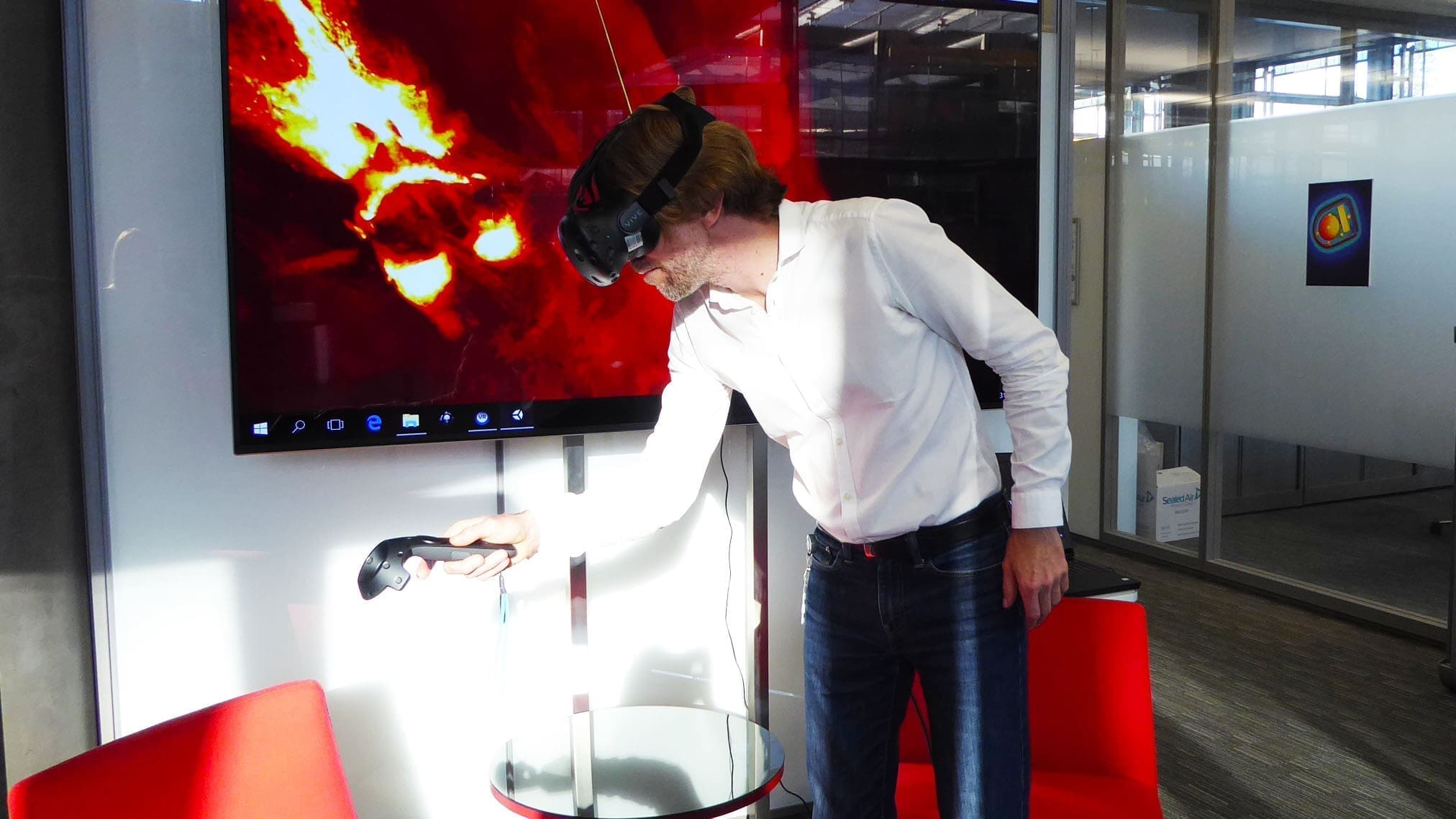
Put your head inside a brain!
A new immersive virtual reality (VR) experience now offers a unique way to visualize and interact with large volumes of 3D anatomical brain data. The system, developed by researchers from the Wyss Center for Bio and Neuroengineering and the University of Geneva, has applications in neurotechnology development, research and surgeon training. A poster describing the system will be presented on Wednesday 15 November at the annual meeting of the Society for Neuroscience 2017, in Washington DC.
Users wearing VR goggles can put their head inside a mouse brain and visualize the 3D structure at cellular resolution. Interactive hand-held pointers allow rapid, natural interaction with the data to highlight, select, slice and zoom.
Play Video
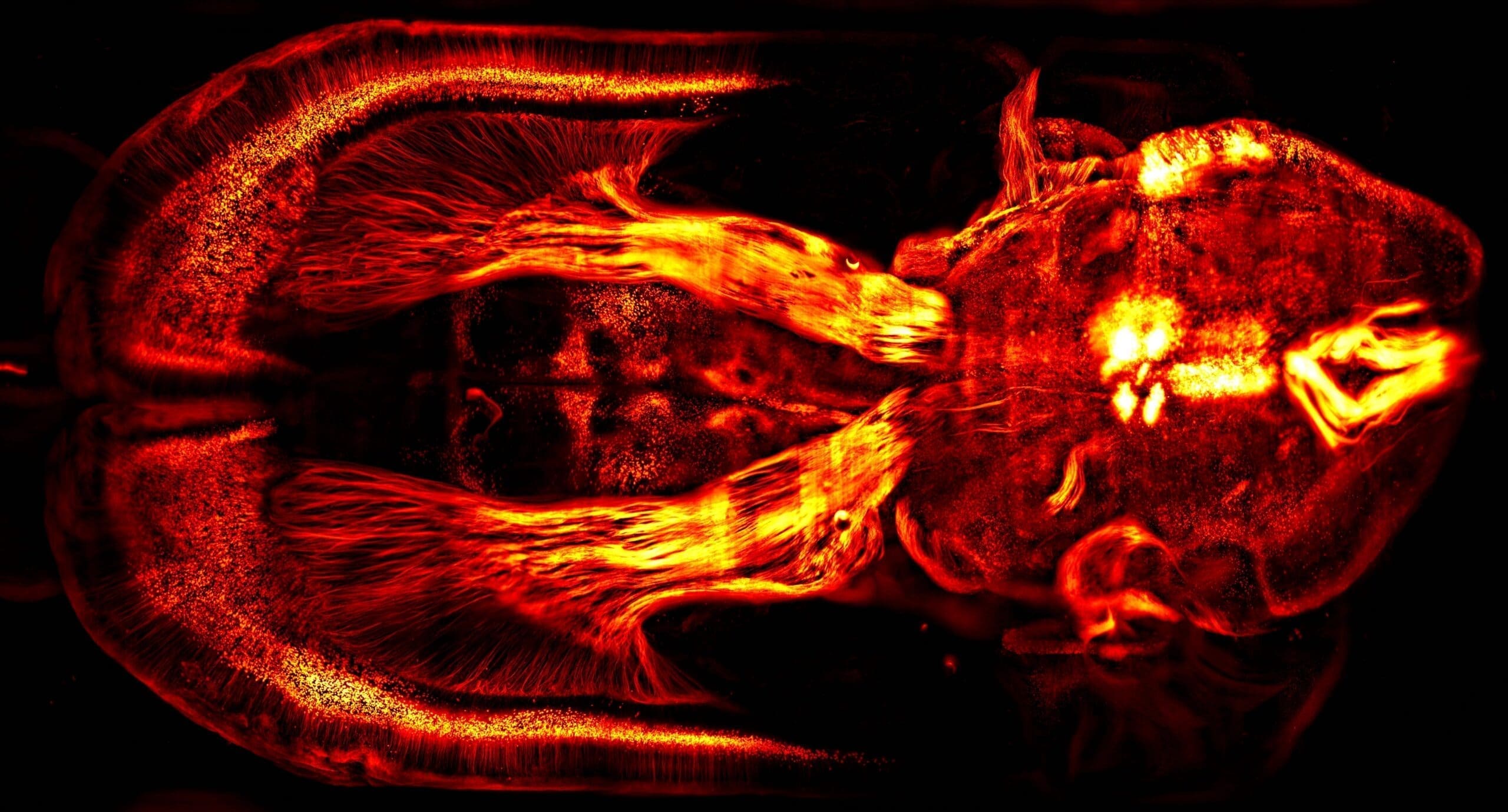
This 3D rotating brain video is part of the VR experience. Brain circuits related to natural reward. Mouse brain injected with a fluorescent anterograde viral tracer placed in the ventral tegmental area to reveal dopaminergic projections, widely implicated in drug and natural reward circuitry. Credit: Christian Lüscher Lab, UNIGE
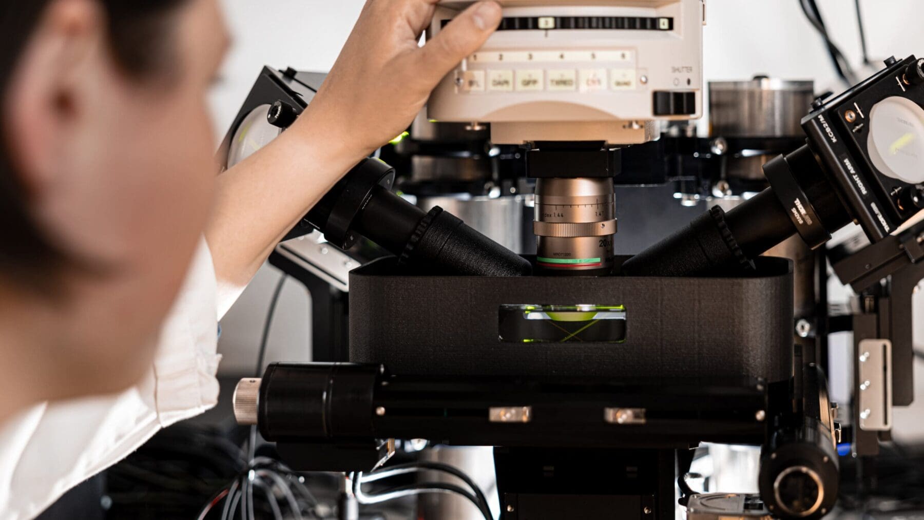
Developed to help handle the huge volumes of data now routinely produced by a new generation of high-resolution imaging techniques, the system is a novel tool to analyze data like that produced by the Wyss Center’s custom lightsheet microscope at Campus Biotech in Geneva, Switzerland.
Dr Stéphane Pages, Staff Scientist at the Wyss Center and Senior Research Associate at the University of Geneva, who managed the microscope fabrication and is lead author of the paper said: “The immense data volumes produced by today’s high-performance microscopes are driving the development of new methods to visualize the brain. We have developed this virtual reality system to reconstruct cellular level neuroanatomical data in 3D space. The system provides a practical solution to experience, analyze and quickly understand these exquisite, high-resolution images.”
Dr Gilles Reymond, Staff Engineer at the Wyss Center said: “In the future, this could prove a very useful tool to gain a new 3D perspective on the complex mechanical and biological interactions between the human brain and new types of MRI compatible brain probes that can be used to help people overcome nervous system disorders.
“It could also be used to help surgeons visualize and practise the steps involved in a complex surgery in virtual reality before performing it on a patient.”
One of only three in the world, the Wyss Center’s lightsheet microscope can image individual neurons – five times thinner than a human hair. The latest viral tracing reveals the distribution of neuronal pathways and even dendritic spines, the micron sized protrusions that are communication points between neurons. Lightsheet microscope imaging is also a valuable tool to visualize and understand the detailed mechanical and biological interactions between novel neural implants and the brain, which is a focus of the Wyss Center’s translational neurotechnology program.
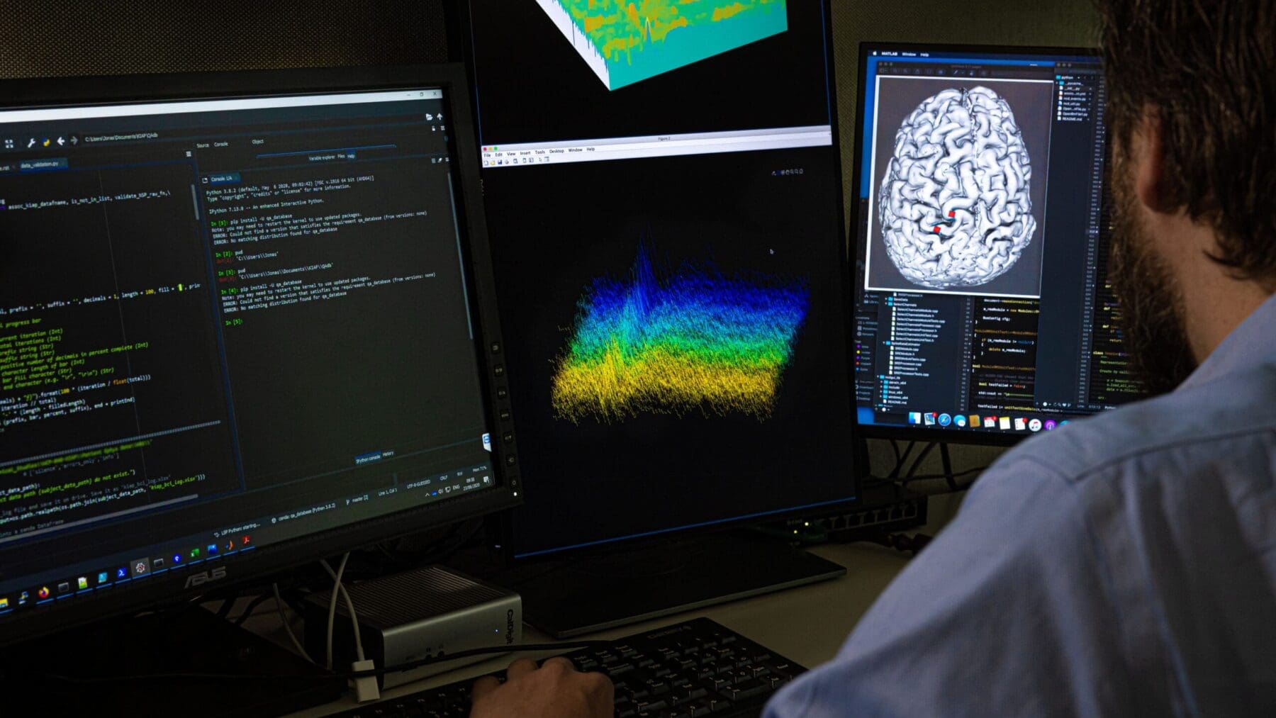
The VR system can also be readily combined with semi-automatic data analysis tools to accelerate the analysis of imaging data and simplify the identification of structures that can be difficult to recognize on a screen.
This 2D brain image captured with a lightsheet microscope is representative of the type of data the VR system can explore in more detail. It shows a mouse brain injected with a fluorescent retrograde virus in the brain stem to reveal labelling of multiple brain wide projections. Credit: Courtine Lab, EPFL (Leonie Asboth and Elodie Rey)
A new model of cooperation and collaboration to accelerate translational research and development in the field of neurotechnology and artificial intelligence.